Welcome to DUTTA Cryo-EM Lab
Cryo-EM Structural Biology lab
Our group investigates the structural and functional mechanisms of bacterial secretion systems, efflux pumps in bacteria, pore-forming toxins, and their interactions with cognate receptors using cryo-electron microscopy (cryo-EM).
A major focus of our research is to characterize liposomes using cryo-EM and to visualize liposome–protein interactions through cryo-EM and cryo-electron tomography (cryo-ET). We also study bacterial toxins and virulence factors in the context of their interactions with host cell membranes—such as those derived from erythrocytes, neutrophils, and macrophages—and their binding to specific cell-surface receptors.
In parallel, we engage in structure-based drug design, developing inhibitory peptides, small-molecule compounds, and antibodies to target these pathogenic systems. Within our laboratory, we integrate multiple complementary techniques, including:
Protein expression and purification, Biochemical and biophysical characterization, Enzyme kinetics and protein chemistry, Fluorescence and confocal imaging, Cell culture studies, Cryo-EM structure determination and cryo-ET, Molecular modeling and simulations for drug design
Overall, our goal is to understand host–pathogen interactions in diseases caused by pathogens such as Staphylococcus aureus and Mycobacterium tuberculosis (Mtb), and to leverage this understanding to develop novel therapeutic strategies to combat these infections.
Recent achievements


.jpeg)
Chatterjee et al. published Nature Communications in 2025. Group resolved PFT with liposomes
Fernando et al. published RSC ADVANCES in 2025. Group resolved PFT with liposomes
Fellow of Electron Microscope Society of India (EMSI), India award for the Year 2024


Science Advances 2022 Bandyopadhyay & Pramanick et al.
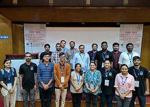
EMSI pre-conference cryo-EM workshop and EMSI international conference July 2025

EMBO CEM3DIP cryo-EM course 2024

Nature Chemical Biology volume
18, pages 1046–1055 (2022)

First high-resolution cryo-EM structure of a pore-forming toxin (PFT) embedded in liposomes was reported in J Cell Biol (2021) 220 (12): e202102035.
RESEARCH INTEREST
1. Metabolic pathways of Mycobacterium tuberculosis
2. Pore-forming toxins and host–pathogen interaction
3. Bacterial secretion systems to understand the secretion of virulence factors.

Structural and functional characterization of the metabolic pathways of Pathogenic bacteria
Mycobacterium tuberculosis (Mtb) is the causative agent of one of the deadliest infectious diseases known to man, tuberculosis. Mtb has a remarkable ability to persist inside the oxidatively hostile environment of human phagocytes encountering Reactive Oxygen Species, Reactive Nitrogen Species, low pH and nutrient starvation etc. This remarkable ability of Mtb made it a successful human pathogen. There are certain products generated from different metabolic pathways of Mtb helps it to combat hostile environment inside human. We are interested to know the mechanism as well as the structural details of proteins present in these kinds of the metabolic pathway.
PUBLICATIONS
Research Publications in International Journals 2016-2025 (Associate Professor)
-
Structural insights into pre-pore intermediates of alpha-hemolysin in the lipidic environment. Chatterjee A, Roy A, Satheesh T, Das PP, Mondal B, Kishore P, Ganji M, Dutta S. Nat Commun. 2025 Jul 10;16(1):6348. doi: 10.1038/s41467-025-61741-x. PMID: 40640160.
-
Pore Formation by Pore Forming Proteins in Lipid Membranes: Structural Insights Through Cryo-EM. Chatterjee A, Naskar P, Mishra S, Dutta S. J Membr Biol. 2025 Aug;258(4):305-322. doi: 10.1007/s00232-025-00344-5. Epub 2025 Mar 28.PMID: 40155553 Review.
-
Cryo-EM reveals conformational variability in the SARS-CoV-2 spike protein RBD induced by two broadly neutralizing monoclonal antibodies. Rencilin CF, Chatterjee A, Ansari MY, Deshpande S, Mukherjee S, Singh R, Jayatheertha SB, Reddy PM, Hingankar N, Varadarajan R, Bhattacharya J, Dutta S. RSC Adv. 2025 May 6;15(18):14385-14399. doi: 10.1039/d5ra00373c. eCollection 2025 Apr 28.PMID: 40330036. Corresponding author.
-
Multiscale Materials Engineering via Self-Assembly of Pentapeptide Derivatives from SARS CoV E Protein. Sarkar D, Khan AH, Polepalli S, Sarkar R, Das PK, Dutta S, Sahoo N, Bhunia A. Small. 2024 Nov;20(45):e2404373. doi: 10.1002/smll.202404373. Epub 2024 Jul 16.PMID: 39011730. Corresponding author.
-
Cryo-EM Reveals the Mechanism of DNA Compaction by Mycobacterium smegmatis Dps2.Garg P, Satheesh T, Ganji M, Dutta S. J Mol Biol. 2024 Nov 1;436(21):168806. doi: 10.1016/j.jmb.2024.168806. Epub 2024 Sep 28.PMID: 39349276 . Corresponding author.
-
Cryo-EM research in India. Shukla AK, Banerjee M, Singh AK, Penmatsa A, Dutta S, Anand R, Sirajuddin M, Srivastava SS. Structure. 2024 Feb 1;32(2):113-119. doi: 10.1016/j.str.2024.01.002.PMID: 38306986
-
Neutralizing Efficacy of Encapsulin Nanoparticles against SARS-CoV2 Variants of Concern.Khaleeq S, Sengupta N, Kumar S, Patel UR, Rajmani RS, Reddy P, Pandey S, Singh R, Dutta S, Ringe RP, Varadarajan R. Viruses. 2023 Jan 25;15(2):346. doi: 10.3390/v15020346.PMID: 36851560
-
Cryo-EM reveals the membrane-binding phenomenon of EspB, a virulence factor of the Mycobacterial Type VII secretion system. Sengupta N, P S, Dutta S. J Biol Chem. 2023 Mar 6:104589. doi: 10.1016/j.jbc.2023.104589. Epub ahead of print. PMID: 36889587. Corresponding author.
-
Cryo-EM-based structural insights into supramolecular assemblies of γ-hemolysin from S. aureus reveal the pore formation mechanism. Mishra S, Roy A, Dutta S. Structure. 2023 Mar 27:S0969-2126(23)00085-0. doi: 10.1016/j.str.2023.03.009. Corresponding author.
-
Kumar S, Singh S, Chatterjee A, Bajpai P, Sharma S, Katpara S, Lodha R, Dutta S, Luthra K. Recognition determinants of improved HIV-1 neutralization by a heavy chain matured pediatric antibody. iScience. 2023 Aug 9;26(9):107579. doi: 10.1016/j.isci.2023.107579. Corresponding author.
-
Glu289 residue in the pore-forming motif of Vibrio cholerae cytolysin is important for efficient b-barrel pore formation. Mondal AK, Sengupta N, Singh M, Biswas R, Lata K, Lahiri I, Dutta S, Chattopadhyay K. J Biol Chem. 2022 Aug 30:102441. doi: 10.1016/j.jbc.2022.102441.
-
Structural insights into thermostable direct hemolysin of Vibrio parahaemolyticus using single-particle cryo-EM. Mishra S, Kundu N, Pramanick I, Kumar A, Chattopadhyay K, Dutta S. Proteins. 2023 Feb;91(2):137-146. doi: 10.1002/prot.26416. Epub 2022 Sep 8. Corresponding author.
-
S-Adenosylmethionine-responsive cystathionine β-synthase modulates sulfur metabolism and redox balance in Mycobacterium tuberculosis. Bandyopadhyay P, Pramanick I, Biswas R, Ps S, Sreedharan S, Singh S, Rajmani RS, Laxman S, Dutta S, Singh A.Sci Adv. 2022 Jun 24;8(25):eabo0097. doi: 10.1126/sciadv.abo0097. Epub 2022 Jun 24.PMID: 35749503. Corresponding author.
-
User-friendly, High-throughput, and Fully Automated Data Acquisition Software for Single-particle Cryo-electron Microscopy. Kumar A, P S, Gulati S, Dutta S.J Vis Exp. 2021 Jul 29;(173). doi: 10.3791/62832.PMID: 34398142. Corresponding author.
-
Single-particle cryo-EM reveals conformational variability of the oligomeric VCC β-barrel pore in a lipid bilayer. Sengupta N, Mondal AK, Mishra S, Chattopadhyay K, Dutta S. J Cell Biol. 2021 Dec 6;220(12):e202102035. doi: 10.1083/jcb.202102035. Epub 2021 Oct Corresponding author.
-
Conformational flexibility and structural variability of SARS-CoV2 S protein. Pramanick I, Sengupta N, Mishra S, Pandey S, Girish N, Das A, Dutta S. Structure. 2021 Apr 26:S0969-2126(21)00122-2. doi: 10.1016/j.str.2021.04.006. Online ahead of print.PMID: 33932324. Corresponding author.
-
A dimeric proteomimetic prevents SARS-CoV-2 infection by dimerizing the spike protein. Khatri B, Pramanick I, Malladi SK, Rajmani RS, Kumar S, Ghosh P, Sengupta N, Rahisuddin R, Kumar N, Kumaran S, Ringe RP, Varadarajan R, Dutta S, Chatterjee J. Nat Chem Biol. 2022 Jun 2. doi: 10.1038/s41589-022-01060-0. Epub ahead of print. PMID: 35654847. Corresponding author.
-
Simplified Approach for Preparing Graphene Oxide TEM Grids for Stained and Vitrified Biomolecules. Kumar A, Sengupta N, Dutta S.Nanomaterials (Basel). 2021 Mar 5;11(3):643. doi: 10.3390/nano11030643. Corresponding author.
-
Multiple nanocages of a cyanophage small heat shock protein with icosahedral and octahedral symmetries. Biswas S, Garg P, Dutta S, Suguna K.Sci Rep. 2021 Oct 25;11(1):21023. doi: 10.1038/s41598-021-00172-2.PMID: 34697325 Corresponding author.
-
Malladi SK, Singh R, Pandey S, Gayathri S, Kanjo K, Ahmed S, Khan MS, Kalita P, Girish N, Upadhyaya A, Reddy P, Pramanick I, Bhasin M, Mani S, Bhattacharyya S, Joseph J, Thankamani K, Raj VS, Dutta S, Singh R, Nadig G, Varadarajan R. Design of a highly thermotolerant, immunogenic SARS-CoV-2 spike fragment. J Biol Chem. 2020 Nov 5:jbc.RA120.016284. doi: 10.1074/jbc.RA120.016284.
-
Comparative Immunogenicity of Bacterially Expressed Soluble Trimers and Nanoparticle Displayed Influenza Hemagglutinin Stem Immunogens. Kar U, Khaleeq S, Garg P, Bhat M, Reddy P, Vignesh VS, Upadhyaya A, Das M, Chakshusmathi G, Pandey S, Dutta S, Varadarajan R. Front Immunol. 2022 May 26;13:890622. doi: 10.3389/fimmu.2022.890622. eCollection 2022.
-
Protective Efficacy of Recombinant Influenza Hemagglutinin Ectodomain Fusions. Mittal N, Sengupta N, Malladi SK, Reddy P, Bhat M, Rajmani RS, Sedeyn K, Saelens X, Dutta S, Varadarajan R. Viruses. 2021 Aug 27;13(9):1710. doi: 10.3390/v13091710.PMID: 34578291
-
N-Terminal Region of Vibrio parahemolyticus Thermostable Direct Hemolysin Regulates the Membrane-Damaging Action of the Toxin. Kundu N, Verma P, Kumar A, Dhar V, Dutta S, Chattopadhyay K.Biochemistry. 2020 Feb 4;59(4):605-614. doi: 10.1021/acs.biochem.9b00937. Epub 2019 Dec 20.
-
Tyrosine in the hinge region of the pore-forming motif regulates oligomeric β-barrel pore formation by Vibrio cholerae cytolysin. Mondal AK, Verma P, Sengupta N, Dutta S, Bhushan Pandit S, Chattopadhyay K.Mol Microbiol. 2020 Oct 21. doi: 10.1111/mmi.14631. Online ahead of print.
-
Malladia SK, Schreiberb D, Pramanicka I, Sridevia MA, Goldenzweigb A, Dutta S, Fleishman SJ, Varadarajan R. One-step sequence and structure-guided optimization of HIV-1 envelope gp140. Current Research in Structural Biology. 2020 Volume 2, Pages 45-55. https://doi.org/10.1016/j.crstbi.2020.04.001.
-
Kundu N ,Verma P ,Kumar A , Dhar V, Dutta S, Chattopadhyay K, N-Terminal Region of Vibrio parahemolyticus Thermostable Direct Hemolysin Regulates the Membrane-Damaging Action of the Toxin. Biochemistry. 2020 Feb 4;59(4):605-614. doi: 10.1021/acs.biochem.9b00937.
-
Ghosh E, Dwivedi H, Baidya M, Srivastava A, Kumari P, Stepniewski T, Kim HR, Lee MH, van Gastel J, Chaturvedi M, Roy D, Pandey S, Maharana J, Guixà-González R, Luttrell LM, Chung KY, Dutta S, Selent J, Shukla AK. Conformational Sensors and Domain Swapping Reveal Structural and Functional Differences between β-Arrestin Isoforms. Cell Rep. 2019 Sep 24;28(13):3287-3299.e6. doi: 10.1016/j.celrep.2019.08.053
-
Datta M, Aroli S, Karmakar K, Dutta S, Chakravortty D, Varshney U. Development of mCherry tagged UdgX as a highly sensitive molecular probe for specific detection of uracils in DNA. Biochem Biophys Res Commun. 2019 Oct 8;518(1):38-43. doi: 10.1016/j.bbrc.2019.08.005. Epub 2019 Aug 9.
-
Koehl A, Hu H, Feng D, Sun B, Zhang Y, Robertson MJ, Chu M, Kobilka TS, Laermans T, Steyaert J, Tarrasch J, Dutta S, Fonseca R, Weis WI, Mathiesen JM, Skiniotis G, Kobilka BK. Structural insights into the activation of metabotropic glutamate receptors. Nature. 2019 Feb;566(7742):79-84.
-
Bhandari S, Biswas S, Chaudhary A, Dutta S, Suguna K. Dodecameric structure of a small heat shock protein from Mycobacterium marinum M. Proteins. 2019 Jan 11. doi: 10.1002/prot.25657.
-
Kumar S, Panda H, Makhdoomi MA, Mishra N, Safdari HA, Chawla H, Aggarwal H, Reddy ES, Lodha R, Kumar Kabra S, Chandele A, Dutta S, Luthra K. An HIV-1 broadly neutralizing antibody from a clade C infected pediatric elite neutralizer potently neutralizes the contemporaneous and autologous evolving viruses. J Virol. 2018 Nov 14. pii: JVI.01495-18. doi: 10.1128/JVI.01495-18.
-
Safdari, HA, Pandey, S, Shukla, AK, Dutta, S. Illuminating GPCR Signaling by Cryo-EM. Trends Cell Biol, 2018, 28, 591-594. Corresponding author.
-
Kesavardhana S, Das R, Datta CM, Ecto R, Srilatha L, DiStefano NS, Swoyer RD, Joyce JG, Dutta S et al. Structure-based Design of Cyclically Permuted HIV-1 gp120 Trimers That Elicit Neutralizing Antibodies. J Biol Chem, 2017, 292, 278-291.
Research Publications in International Journals 2010-2016 (Ph.D. & Postdoctoral Fellow)
-
Yee A, Oleskie AN, Dosey AM, Kretz CA, Gildersleeve RD, Dutta S, Su M, Ginsburg D, Skiniotis G. Visualization of an N-terminal fragment of von Willebrand factor in complex with factor VIII. Blood. 2015, 126:939-42.
-
Xu K, Chan YP, Bradel-Tretheway B, Akyol-Ataman Z, Zhu Y, Dutta S, Yan L, Feng Y, Wang LF, Skiniotis G, Lee B, Zhou ZH, Broder CC, Aguilar HC, Nikolov DB. PLoS Pathog. Crystal Structure of the Pre-fusion Nipah Virus Fusion Glycoprotein Reveals a Novel Hexamer-of-Trimers Assembly. 2015 8;11(12).
-
Dutta, S.*, Whicher, J. R.*, Hansen, D. A., Hale, W. A., Chemler, J. A., Congdon, G. R., Narayan, A. R., Håkansson, K., Sherman, D. H., Smith, J. L., and Skiniotis, G. Structure of a modular polyketide synthase. Nature. 2014, 510:512-17. [*Equal contribution].
-
Whicher, J. R.*, Dutta, S.*, Hansen, D. A., Hale, W. A., Chemler, J. A., Congdon, G. R., Narayan, A. R., Håkansson, K., Sherman, D. H., Smith, J. L., and Skiniotis, G. Structural rearrangements of a polyketide synthase module during its catalytic cycle. Nature. 2014, 510:560-64. [*Equal contribution].
-
Akey DL, Brown WC, Dutta S, Konwerski J, Jose J, Jurkiw TJ, DelProposto J, Ogata CM, Skiniotis G, Kuhn RJ, Smith JL. Flavivirus NS1 structures reveal surfaces for associations with membranes and the immune system. Science. 2014, 343 :881-85.
-
Dutta, S, Banerjee, KK, Ghosh AN. Cryo-electron microscopy reveals the membrane insertion mechanism of V. cholerae hemolysin. J Biomol Struct Dyn. 2013, 32:1434-42.
-
Lyon, A.M., Dutta, S., Skiniotis, G., and Tesmer, J.J.G. Characterization of phospholipase C-β3 complexes by X-ray crystallography and single particle cryo-electron microscopy. Transactions of the American Crystallographic Association (2013).
-
Lyon AM, Dutta S, Boguth CA, Skiniotis G, Tesmer JJ. Full-length Gα(q)-phospholipase C-β3 structure reveals interfaces of the C-terminal coiled-coil domain. Nat Struct Mol Biol. 2013, 20(3):355-62.
-
Chan YP, Lu M, Dutta S, Yan L, Barr J, Flora M, Feng YR, Xu K, Nikolov DB, Wang LF, Skiniotis G, Broder CC. Biochemical, conformational, and immunogenic analysis of soluble trimeric forms of henipavirus fusion glycoproteins. J Virol. 2012, 86(21):11457-71.
-
Westfield GH*, Rasmussen SG*, Su M*, Dutta S*, DeVree BT, Chung KY, Calinski D, Velez-Ruiz G, Oleskie AN, Pardon E, Chae PS, Liu T, Li S, Woods VL Jr, Steyaert J, Kobilka BK, Sunahara RK, Skiniotis G.Structural flexibility of the G alpha s alpha-helical domain in the beta2-adrenoceptor Gs complex. Proc Natl Acad Sci. U S A. 2011, 108(38):16086-91. [* Equal contribution]
-
Gu L, Eisman EB, Dutta S, Franzmann TM, Walter S, Gerwick WH, Skiniotis G, Sherman DH. Tandem acyl carrier proteins in the curacin biosynthetic pathway promote consecutive multienzyme reactions with a synergistic effect. Angew Chem Int Ed Engl. 2011, 50(12):2795-98.
-
Dutta S, Mazumdar B, Banerjee KK, Ghosh AN. Three-dimensional structure of different functional forms of the Vibrio cholerae hemolysin oligomer: a cryo-electron microscopic study. J Bacteriol. 2010, 192(1):169-78.

“Kindness is the language which the deaf can hear and the blind can see”
Mark Twain
Quote

Course & Teaching
Electron microscopy and 3D image processing for Life sciences. (MB 212 JAN 2:0)


NPTEL Course-Fundamentals of Cryo-Electron Microscopy (Cryo-EM) and 3D Image Processing in Structural BiologyBy Prof. Somnath Dutta | IISc Bangalore
https://nptel.ac.in/courses/102108668
Electron microscopy and 3D image processing for Life sciences. (MB 222 JAN 3:0)
UB207 General Biochemistry
Previous Course
Transmission Electron Microscopy & Cryo-EM and single particle analysis. A 2-month (50 hours) hands-on training program, 2021-2024. ~40 students.
Electron microscopy and 3D image processing for Life sciences. (MB 212 JAN 2:0). Academic year 2016-2022
Advance course on Biological Cryo-EM sample preparation and 3D image reconstruction (2:1)
.jpeg)
Group Leader
Dr. Somnath Dutta
Associate Professor
Ph.D. from NICED, Kolkata, 2010
Post Doc. Skiniotis Lab, University of Michigan, Michigan, Ann Arbor, USA From 2010-2016
Assistant Profesor, MBU, IISc, Bangalore 2016-2022.
Associate Professor, MBU, IISc, Bangalore 2022-

Cryo-EM Facility Manager
Drose Ignatious Shane
I completed both my Bachelor’s and Master’s degrees in Biotechnology from the Kalasalingam Academy of Research and Education, Virudhunagar, Tamil Nadu. I had the privilege of carrying out my master’s thesis at the Indian Institute of Science (IISc), Bangalore, under the guidance of Dr. Somnath Dutta, where I developed a strong foundation in structural biology and Cryo-Electron Microscopy (Cryo-EM). Currently, I serve as the Facility Manager at the Advanced Centre for Cryo-Electron Microscopy, IISc, where I support and collaborate with researchers in high-resolution structural studies. My work focuses on pushing the frontiers of Cryo Single Particle Analysis (Cryo-SPA) and Cryo-Electron Tomography (Cryo-ET).

Ph.D student
Surekha Padmanaban
I did my bachelor’s in biotechnology from Rajalakshmi Engineering College, Chennai, Tamil Nadu. Followed by master’s in medical biotechnology from Indian Institute of Technology, Hyderabad, Telangana. My interest in structural studies of macromolecular complexes has made me join Dr. Somnath’s lab in molecular biophysics department as a Ph.D student in August 2019.
surekhap@iisc.ac.in

Int. Ph.D. student
Bapan Mondal
Integrated Ph.D. student
Completed B.Sc. in Zoology (2019-22) from Ramakrishna Mission Vidyamandira, Howrah.

Int. PhD Student
Partho Pratim Das
Integrated PhD Student
Done Under graduation(2019-2022) from Ramakrishna Mission Vidyamandira, Belur


Ph D Student
Dipanwita Mandal
I did my B.Tech in biotechnology from the National Institute of technology Durgapur. After that I joined MBU for my PhD degree. My research work is mainly focussed on Mycobacterium tuberculosis secretion system.
Postdoctoral Research fellow
Dr. Nitin Srivastava
I received my PhD in Biochemistry and Molecular Biology from the Chemistry Department of the Indian Institute of Technology (IIT) Delhi. I further pursued a post-doctoral research position in Microbial genetics at the Department of Biotechnology and Biochemical Engineering at IIT Delhi. Both my PhD and post-doctoral tenure focused on various microbial enzymes, their engineering, biochemical and biophysical characteristics, and biotechnological/pharmaceutical applications. Currently, I’m working as a Research Associate at the Advanced Centre for Cryo-Electron Microscopy Facility in Dr Somnath Dutta's group at MBU-IISc, where I am involved in the field of Cryo-Electron Microscopy to gain an understanding of the structural overviews of human metabolic transsulfuration enzymes and apicomplexan perforin-like proteins using Cryo-EM.

Ph.D. Student
Clayton Fernando R
I received my B. Tech and M.Tech degree in Biotechnology from Kalasalingam Academy of Research and Education and did my research projects in various premier research institutions such as CSIR-IICB, IISc, and Aravind Medical Research Foundation and published some research papers. Also, two times I'm selected as an INSA-NASI-IASc summer research fellow. Currently, I wish to explore macromolecular complexes involved in pathogenesis using the pioneering structural biology tool, Cryo-EM and will assist in the development of drugs and vaccines. In the same way, I'm maintaining the Cryo-EM facility.

PhD Student
Sainath Polepalli
I completed my Bachelor of Technology (B.Tech) in Industrial Biotechnology from Alagappa College of Technology (ACTech), Anna University, Chennai (2016-2020). I am interested in learning the Protein Structure elucidation techniques. I joined Dr. Somanth Dutta’s lab in MBU as a Ph.D. student in 2020 to gain experience in Protein Structure determination using the Cryo-EM.

Ph.D. Student
Arnab Chatterjee A
MSc-PhD student
#CryoEM Former Undergraduate student at Banaras Hindu University

PhD student
Shreya Suman
Molecular Biophysics Unit

Int. PhD student
Subhajit Mahanty
Molecular Biophysics Unit

PhD student
Asmita Roy
Molecular Biophysics Unit

UG student
Anurag Majumdar
Molecular Biophysics Unit
Lab News
-
Arnab Chatterjee will present his recent work in ETOX 2023, EUROPEAN WORKSHOP ON BACTERIAL PROTEIN TOXINS organised by University of Dundee UK and University of Sheffield, UK.
-
Nayanika Sengupta will present her recent work in Mechanisms of Membrane Transport (GRS), Gordon Research Seminar in June 17 - 18, 2023 at Eurotel Victoria, Les Diablerets, VD, Switzerland.
-
-
https://www.msn.com/en-in/news/other/iisc-study-shows-forms-of-sars-cov2-s-protein/ar-AAKAq3N
-
https://www.iisc.ac.in/novel-insights-into-the-structure-of-sars-cov-2-spike-protein/
-
https://biopatrika.com/2021/06/03/interview-conformational-flexibility-and-structural-variability-of-sars-cov2-s-protein/
-
https://www.ibtimes.co.in/breakthrough-iiscs-study-gives-new-insights-into-structure-covid-spike-protein-837713
-
7. http://ianslive.in/news/iisc_s_study_gives_new_insights_into_structure_of_covid_spike_protein-793774/NATION/1/google
-
https://react.etvbharat.com/english/national/science-and-technology/iiscs-study-gives-new-insights-into-structure-of-covid-spike-protein/na20210618111653595
-
https://www.ibtimes.co.in/breakthrough-iiscs-study-gives-new-insights-into-structure-covid-spike-protein-837713
Recognizing the enormity of the challenges our society faces, DUTTA Lab (Cryo-EM Structural Biology lab) is dedicated to making a lasting impact through our programs. While our efforts are driven by our organization’s singular focus, we spread a wide net by investing in a variety of progressive strategies. Learn more about our initiatives and get involved yourself.

APMC Meeting Feb 2020
Past Fellows
1. ISHIKA PRAMANICK (July 2016-April 2022). Currently at Norwich BioScience Institutes, Colney, Norwich
2. Anil Kumar (July 2017-April 2021) Cryo-EM Manager. Currently at UW-Madison Cryo-EM Research Center.
3. Alakta Das (May 2017 - July 2021), UG student of IISc – BS & MS Thesis. Currently Darwin Trust Fellow PhD student, University of Edinburgh, Edinburgh, UK.
4. Anuradha Choudhary (Project Fellow) May 2016-May 2018. (Now a Ph.D. student at International Institute of Molecular and Cell Biology, Warsaw, Poland).
5. Haaris Ahsan Safdari (MS student 2017-2019).
6. Abirlal Mukherjee (Project Fellow) 2018-2019. (Now a Ph.D. student at the Indian Institute of Technology, Roorkee).
7. Manish Sarkar (Project Fellow) 2016-2017 (Now Ph.D. Student at Bose Institute, Kolkata).
8. Navyashree V (July-Sep 2017), Bangalore University – Summer training
9. Suresh Kumar (Project Fellow)
10. Akansha Patel (M.Tech Final Year Project Student) 2016-2017
11. Preeti (Postdoc May 2017-May 2018)
12. Ayushi Shukla (Project Fellow)
13. Puja – Summer training
14. Gyana Prakash Mahapatra (Project Assistant)
Contact DUTTA Lab (Cryo-EM Structural Biology lab)
Indian Institute of Science, Molecular Biophysics Unit, Annex building, 1st Floor, Bangalore 560012
080-2293 3453
